- 029-86354885
- 18392009562
0.8nm/1.4nm納米金偶連磷脂DPPE產(chǎn)品使用說明書
西安瑞禧生物科技有限公司推出一款DPPE磷脂通過化學(xué)鍵偶連0.8納米或1.4納米的納米金產(chǎn)品,該產(chǎn)品穩(wěn)定性高,目前銷售的二個包裝規(guī)格6nmol和30nmol。
該產(chǎn)品的使用說明書如下:
PRODUCT INFORMATION
DIPALMITOYLPHOSPHATIDYL ETHANOLAMINE-NANOGOLD®
Product Name: DIPALMITOYLPHOSPHATIDYLETHANOLAMINE-NANOGOLD®
Catalog Number: 4021
Appearance: Dark brown solid
Revision: 1.2 (March 2000)
GENERALINFORMATION:
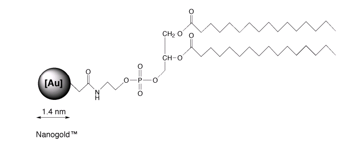
DPPE-NANOGOLD® consists of the 1.4 nmNANOGOLD® particle covalently linked to a single molecule of dipalmitoylphosphatidyl ethanolamine. Conjugation is via the amine group on theethanolamine head of the molecule. It is intended as a lipid label for use withmicelles and other dual-phase systems1,2 in the same manner as the previouslydescribed undecatungstate membrane label3 . It has been used to prepare gold-decoratedliposomes1 , and also to track the dellivery of a liposomally encapsulatedantifungal drug2 . Its structure is shwon in figure 1:

Figure1:Structure of dipalmitoyl phosphatidyl ethanolamine-NANOGOLD®(not shown to scale)
DPPE-NANOGOLD® is supplied as a solid,dried from methanol solution. It should be refrigerated upon receipt, andstored at 2 - 8°C.
INSTRUCTIONSFOR USE:
DPPE-NANOGOLD® is hydrophobic, and caninsert into organic phases in systems such as micelles.1 It is soluble inmethanol and in methanol-trichlrormethane and methanol-dichloromethanemixtures. Extinction coefficients at specific wavelengths are given below formethanol solution:
WAVELENGTH (nm) EXTINCTION COEFFICIENT*
280 2.25 × 100000
420 1.12 × 100000
Measured for 5 X 10⁻⁶ M solution in methanol.
Heavily gold-labeled liposomes may beformed by dissolving the Nanogold®-DPPE in chloroform, mixing with water,evaporating the chloroform, then using sonication or other usual preparativemethods to form vesicles1 . Nanogold®-DPPE may also be diluted with unlabeledDPPE or with other lipids and used to prepare less heavily labeled liposomes.Incorporation of 0.1 to 1 % Nanogold®-DPPE with an unlabeled lipid is usuallyappropriate for preparing Nanogold®-labeled liposomes with the same propertiesand morphology as those prepared without the gold label2 . Once incorporatedinto micelles or other structures, it may be used according to the proceduresrequired by individual experiments in the same manner as unlabeled DPPE.
GENERALCONSIDERATIONS FOR STAINING WITH NANOGOLD® REAGENTS
Because the 1.4 nm NANOGOLD® particles areso small, over staining with OsO4, uranyl acetate or lead citrate may tend toobscure direct visualization of individual NANOGOLD® particles. Threerecommendations for improved visibility of NANOGOLD® are:
l. Use of reduced amounts or concentrationsof usual stains.
2. Use of lower atomic number stains suchas NANOVAN?, a Vanadium based stain.
3. Enhancement of NANOGOLD® with silverdevelopers, such as LI SILVER or HQ SILVER
TEMPERATURECAUTION:
NANOGOLD® particles are not stable above50°C. Best results are obtained at room temperature or 4°C. Avoid 37°Cincubations. Use low temperature embedding media (e.g., Lowicryl) if labelingbefore embedding; do not bake tissue blocks with NANOGOLD®. If your experimentrequires higher temperature embedding, then silver enhancement should beperformed before embedding.
THIOLCAUTION:
NANOGOLD® particles degrade upon exposureto concentrated thiols such as ?-mercaptoethanol or dithiothreitol. If suchreagents must be used, concentrations should be kept below 1 mM and exposurerestricted to 10 minutes or less.
MICROSCOPE
For most work, silver enhancement isrecommended to give a good signal in the electron microscope (see below). Forparticular applications, visualization of the NANOGOLD® directly may bedesirable. Generally this requires very thin sampl;es and precludes the use ofother stains.
NANOGOLD® provides a much improvedresolution and smaller probe size over other colloidal gold antibody products.However, because NANOGOLD® is only 1.4 nm in diameter, it will not only besmaller, but will appear less intense than, for example, a 5 nm gold particle.With careful work, however, NANOGOLD® may be seen directly through thebinoculars of a standard EM even in 80 nm thin sections. However, achieving thehigh resolution necessary for this work may require new demands on your equipmentand technique. Several suggestions follow:
1. Before you start a project withNANOGOLD® it is helpful to see it so you know what to look for. Dilute theNANOGOLD® stock 1:5 and apply 4 μl to a grid for 1 minute. Wick the drop andwash with deionized water 4 times.
2. View NANOGOLD® at 100,000 Xmagnification with 10 X binoculars for a final magnification of 1,000,000 X.Turn the emission up full and adjust the condenser for maximum illumination.
3. The alignment of the microscopeshould be in order to give 0.3 nm resolution. Although the scope should be wellaligned, you may be able to skip this step if you do step 4.
4. Objective stigmators must beoptimally set at 100,000 X. Even if the rest of the microscope optics are notperfectly aligned, adjustment of the objective stigmators may compensate andgive the required resolution. You may want to follow your local protocol forthis alignment but since it is important, a brief protocol is given here:
a. At 100,000 X (1 X 106 with binoculars), over focus, under focus, then setthe objective lens to in focus. This is where there is the least amount ofdetail seen.
b. Adjust each objective stigmator to give the least amount of detail in theimage.
c. Repeat steps a and b until the in focus image contains virtually no contrast,no wormy details, and gives a flat featureless image.
5. Now underfocus slightly, moveto a fresh area, and you should see small black dots of 1.4 nm size. This isthe NANOGOLD®. For the 1:5 dilution suggested, there should be about 5 to 10gold spots on the small viewing screen used with the binoculars. Contrast andvisibility of the gold clusters is best at 0.2 - 0.5 m defocus, and is muchworse at typical defocus values of 1.5 - 2.0 m commonly used for proteinmolecular imaging.
6. In order to operate at highmagnification with high beam current, thin carbon film over fenestrated holeyfilm is recommended. Alternatively, thin carbon or 0.2% Formvar over a 1000mesh grid is acceptable. Many plastic supports are unstable under theseconditions of high magnification/high beam current and carbon is thereforepreferred. Contrast is best using thinner films and thinner sections.
7. Once you have seen NANOGOLD®you may now be able to reduce the beam current and obtain better images onfilm. For direct viewing with the binoculars reduction in magnification from1,000,000 X to 50,000 X makes the NANOGOLD® much more difficult to observe andnot all of the golds are discernable. At 30,000 X (300,000 X with 10 Xbinoculars) NANOGOLD® particles are not visible. It is recommended to view at1,000,000 X, with maximum beam current, align the objective stigmators, andthen move to a fresh area, reduce the beam, and record on film.
8. If the demands of highresolution are too taxing or your sample has an interfering stain, a very goodresult may be obtained using silver enhancement to give particles easily seenat lower magnification.
SILVERENHANCEMENT OF NANOGOLD® FOR EM:
NANOGOLD® will nucleate silver depositionresulting in a dense particle 2-80 nm in size or larger depending ondevelopment time. If specimens are to be embedded, silver enhancement isusually performed after embedding, although it may be done first. It must becompleted before any staining reagents such as osmium tetroxide, lead citrateor uranyl acetate are applied, since these will nucleate silver deposition inthe same manner as gold and produce non-specific staining. With NANOGOLD®reagents, low-temperature resins (eg Lowicryl) should be used and the specimenskept at or below room temperature until after silver development has beencompleted. Silver development is recommended for applications of NANOGOLD® inwhich these stains are to be used, otherwise the NANOGOLD® particles may bedifficult to visualize against the stain.
Our LI SILVER silver enhancement system isconvenient and not light sensitive, and suitable for all applications. Improvedresults in the EM may be obtained using HQ SILVER, which is formulated to giveslower, more controllable particle growth and uniform particle size distribution.
Specimens must be thoroughly rinsed withdeionized water before silver enhancement reagents are applied. This is becausethe buffers used for antibody incubations and washes contain chloride ions andother anions which form insoluble precipitates with silver. These are oftenlight-sensitive and will give non-specific staining. To prepare the developer,mix equal amounts of the enhancer and initiator immediately before use.NANOGOLD® will nucleate silver deposition resulting in a dense particle 2-20 nmin size or larger depending on development time. Use nickel grids (not copper).
The relevent procedure for immunolabelingshould be followed up to step 7 as described above. Silver enhancement is thenperformed as follows:
1. Rinse with deionized water (2 X5 mins).
2. OPTIONAL (may reducebackground): Rinse with 0.02 M sodium citrate buffer, pH 7.0 (3 X 5 mins).
3. Float grid with specimen onfreshly mixed developer for 1-8 minutes, or as directed in the instructions forthe silver reagent. More or less time can be used to control particle size. Aseries of different development times should be tried, to find the optimum timefor your experiment. With HQ silver, a development time of 6 min. gives 15-40nm round particles.
4. Rinse with deionized water (3 X1 min).
5. Mount and stain as usual.
Fixing with osmium tetroxide may cause someloss of silver; if this is found to be a problem, slightly longer developmenttimes may be appropriate.
PRODUCT INFORMATION
Product Name: DIPALMITOYLPHOSPHATIDYLETHANOLAMINE-UNDECAGOLD
Catalog Number: 4023
Appearance: Yellow-orange solid
Revision: 1.1 (March 2000)
GENERAL INFORMATION
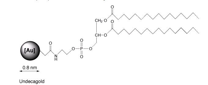
UNDECAGOLD is the smallest gold labelavailable, prepared using a discrete gold compound rather than a colloid.1DPPEUNDECAGOLD consists of the 0.8 nm UNDECAGOLD particle covalently linked toa single molecule of dipalmitoyl phosphatidyl ethanolamine. Conjugation is viathe amine group on the ethanolamine head of the molecule. It is intended as alipid label for use with micelles and othe dual-phase systems. Its structure isshwon in figure 1:

INSTRUCTIONSFOR USE
DPPE-UNDECAGOLD is hydrophobic, and caninsert into organic phases in systems such as micelles. It is slightly solublein methanol and in methanol-trichloromethane and methanol-dichloromethanemixtures. Once incorporated into micelles or other structures, it may be usedaccording to the procedures required by individual experiments in the samemanner as unlabeled DPPE.
WAVELENGTH (nm) EXTINCTION COEFFICIENT*
420 0.471 X 100000
Measured for 5 X 10⁻⁶ M solution inmethanol.
| 序號 | 新聞標(biāo)題 | 瀏覽次數(shù) | 作者 | 發(fā)布時間 |
|---|---|---|---|---|
| 1 | 抗氧化小分子70831-56-0,菊苣酸Cichoric Acid,6537-80-0的制備過程 | 751 | 瑞禧生物 | 2023-03-30 |
| 2 | 活性氧ROS小分子Dapsone,cas:80-08-0,氨苯砜的制備過程-瑞禧科研 | 667 | 瑞禧生物 | 2023-03-30 |
| 3 | HBPS-N3,Azide-PEG-HBPS,疊氮化超支化聚苯乙烯高分子聚合物的制備過程 | 779 | 瑞禧生物 | 2023-03-17 |
| 4 | l-PS-PhN3,Azide疊氮Azido偶聯(lián)線性聚苯乙烯雙鏈的制備過程 | 723 | 瑞禧生物 | 2023-03-17 |
| 5 | N3-PS-N3,Azido-PS-Azido/Azide,雙疊氮官能團(tuán)修飾聚苯乙烯的制備方法 | 682 | 瑞禧生物 | 2023-03-17 |
| 6 | PS-N3,Azido-PS,疊氮Azide修飾聚苯乙烯/高分子聚合物的制備過程 | 853 | 瑞禧生物 | 2023-03-17 |
| 7 | Azido-PEG2-t-Butylester/1271728-79-0,疊氮N3/ZAD修飾叔丁酯化合物的制備方法 | 698 | 瑞禧生物 | 2023-03-14 |


 400-115-0588
 在線咨詢
在線咨詢


 庫存查詢
庫存查詢